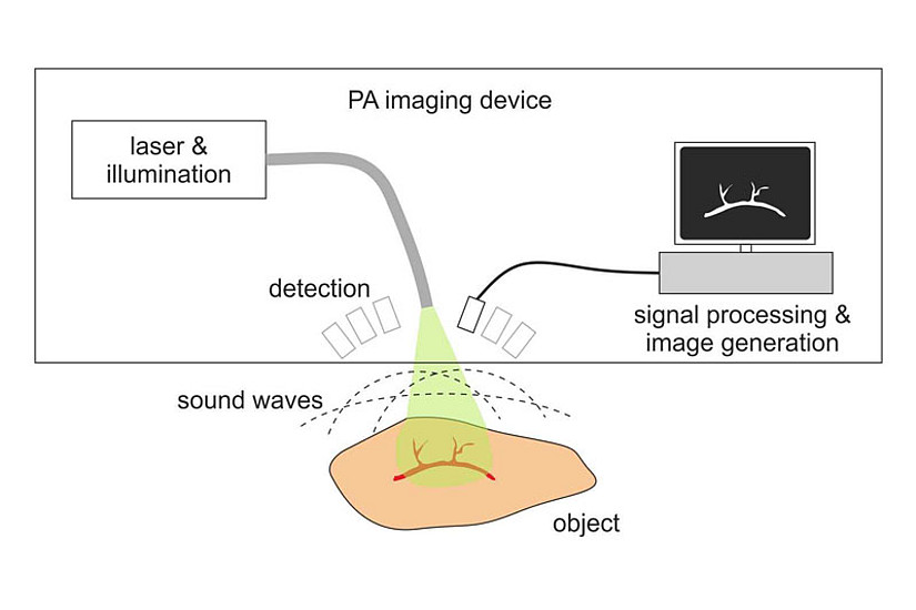
The method of photoacoustic imaging, based on the generation of ultrasound by irradiation with short laser pulses, has seen an enormous boost within the last 20 years. The main goal of this method is to look into biological tissue and to obtain sharp images of tissue structures, mainly the vasculature and the contained blood oxygenation. Implementations range from microscopy of shallow tissue regions to tomography, the latter enabling the imaging of structures as deep as several centimeters with sub-millimeter resolution, despite the diffuse light propagation. Numerous commercial instruments for preclinical research are already available. Currently, intensive developments of devices for clinical use are underway, with a main focus on tomography for the diagnostics of breast cancer.
The photoacoustic group of Guenther Paltauf and Robert Nuster, part of the work group “Optics of Nano and Quantum Materials,” has been involved for many years in research on special methods for ultrasound detection and image reconstruction in photoacoustic tomography and microscopy. Following an invitation by the Editor-in-chief of the “Journal of Applied Physics”, they have authored, together with Martin Frenz from the University of Bern, a “Perspective” article entitled „Progress in biomedical photoacoustic imaging instrumentation towards clinical application“ (https://doi.org/10.1063/5.0028190). In this paper, they give first an overview of recent developments related to instrumentation and image generation. This is followed by an outlook on possible future advances with the goal to take advantage of the unique properties of photoacoustic imaging in medical diagnostics. This includes the use of large ultrasound arrays for real-time three-dimensional imaging, the miniaturization of endoscopic probes, and the use of machine learning for data processing.
With the anticipated developments, the authors believe that within a couple of years various devices will be available for widespread clinical applications.


